Study 4: Anti-cellulite
Dermatological efficacy study on cellulite skin
Method
- Randomized, double-blind, placebo-controlled study with 20 healthy female volunteers
- Age: 44 ± 9 y, average BMI 25
- Body Serum containing Dermatopoietin® or placebo were applied on either the left or right thigh for 8 weeks.
Measurements
Skin structure, in particular the hypodermal – dermal junction distance, by ultrasonography at 20 MHz on day 0, 28 and 56.
Results
Cellulite skin is characterized by lipid intrusions from the hypodermis into the dermis, resulting in a ‘bumpy’ interface between these two layers and a rough skin surface. Application of the Serum containing Dermatopoietin® led to a flattening of the dermal-hypodermal interface, a reduction of the lipid ‘blobs’ in the dermis, a more dense dermis and in a smoother skin surface as shown below.
Ultrasonograms of cellulite skin
Color coding: The stronger and lighter the color the denser the proteins. Visible are the layers of keratin in the epidermis (yellow band at left) and of collagen in the dermis (wide green layer). The application of Dermatopoietin® leads to a build-up of collagen in the dermis and a strengthening of the epidermis. Black pixels represent proteoglycans, lipids and water.
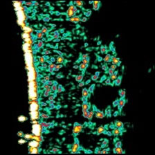
Typical appearance of thigh skin of a 55-year old woman with strong cellulite
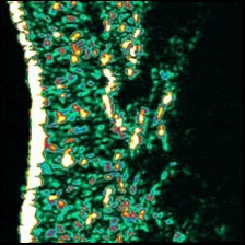
After 4-week application of Dermatopoietin®
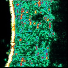
After 8-week application of Dermatopoietin®
Regular photographs
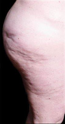
Before Dermatopoietin®
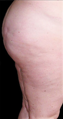
After 8-week application of Dermatopoietin®
A presentation with more detailed information of the study can be found here.